Science
Researchers Unlock High-Resolution Imaging of Cellular Organelles
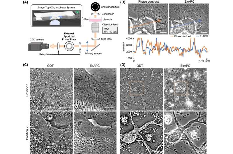
Researchers at the University of Tsukuba have developed a groundbreaking technique for high-resolution imaging of cellular organelles, including nuclei and mitochondria. This advancement, achieved using an external apodized phase contrast (ExAPC) microscope, effectively reduces halo artifacts—misleading images caused by light diffraction. The study, published on November 10, 2025, in The FEBS Journal, demonstrates how this innovative approach allows for the examination of organelle dynamics and spatial organization without the need for labeling.
The study highlights the importance of observing organelles, which are critical for cellular functions. The nucleus stores genetic information, while mitochondria play a vital role in energy production. In addition to these “membranous organelles,” cells also contain “nonmembranous organelles,” which form through molecular assembly. The distribution and morphology of these structures significantly influence cellular fate and function.
Traditionally, fluorescence-based imaging techniques have dominated the field of organelle visualization. However, these methods have notable limitations, such as phototoxicity from intense light exposure and the degradation of fluorescent dyes, which restricts the number of observable structures at one time. To address these challenges, phase contrast microscopy has emerged as a viable alternative, allowing visualization of transparent structures without staining. Yet, conventional phase contrast methods often suffer from halo artifacts that obscure details.
In their recent work, the researchers employed ExAPC microscopy to overcome these limitations. By incorporating an optical mechanism to suppress halo formation, they achieved high-resolution, label-free views of multiple organelles during dynamic cellular processes, including the cell cycle. For the first time, they successfully visualized biomolecular condensate-like structures, the composition of which remains unidentified.
The study observed various cellular behaviors, including the growth of lipid droplets and the processes of mitochondrial fission and fusion. These findings revealed significant heterogeneity in the behavior of individual organelles, while the overall cellular system maintained a remarkable level of order and stability. This robust system is essential for the proper functioning of cells.
The implications of this research are profound. The ExAPC microscopy technique offers a powerful tool for studying biological dynamics in their natural states. It holds significant promise for advancing our understanding of various pathological conditions linked to altered organelle morphology, quantity, and spatial organization. Such conditions include cancer, metabolic disorders, and neurodegenerative diseases. This technology could pave the way for the development of novel diagnostic and therapeutic strategies aimed at addressing these critical health issues.
In summary, the work conducted by the team at the University of Tsukuba marks a significant advancement in cellular imaging technology. By providing a clearer, more accurate view of organelle behavior and organization, it opens up new avenues for research and potential clinical applications.
For further details, refer to the original study: Hiroshi Ohno et al, Label‐free imaging of intracellular structures in living mammalian cells via external apodization phase‐contrast microscopy, The FEBS Journal (2025). DOI: 10.1111/febs.70286.

Goldie Hawn Celebrates Daughter Kate Hudson’s Star Power

Study Reveals Two Harmful Gene Variants Can Restore Function

Dietitians Warn Against Mixing Vitamins with Morning Coffee
Vehicle Crashes into JLC Logistics Building in Rogers, 3 Injured

Trump Administration’s Actions Raise Alarms Over Free Speech
Jayda Cheaves Claims Lil Baby and Ari Fletcher Had an Affair
Rachel Campos-Duffy Exits FOX Noticias; Andrea Linares Steps In
Piper Rockelle Shatters Record with $2.3M First Day on OnlyFans

Meta’s 2026 AI Policy Sparks Outrage Over Privacy Concerns
Leon Goretzka Considers Barcelona Move as Transfer Window Approaches
-
 Entertainment1 day ago
Jayda Cheaves Claims Lil Baby and Ari Fletcher Had an Affair
-
 Top Stories1 month ago
Rachel Campos-Duffy Exits FOX Noticias; Andrea Linares Steps In
-
 Top Stories2 weeks ago
Piper Rockelle Shatters Record with $2.3M First Day on OnlyFans
-
Top Stories2 weeks ago
Meta’s 2026 AI Policy Sparks Outrage Over Privacy Concerns
-
 Sports2 weeks ago
Leon Goretzka Considers Barcelona Move as Transfer Window Approaches
-
 Top Stories2 weeks ago
Urgent Update: Denver Fire Forces Mass Evacuations, 100+ Firefighters Battling Blaze
-
 Health2 months ago
Terry Bradshaw Updates Fans on Health After Absence from FOX NFL Sunday
-
 Top Stories2 weeks ago
OnlyFans Creator Lily Phillips Reconnects with Faith in Rebaptism
-
 Sports2 weeks ago
South Carolina Faces Arkansas in Key Women’s Basketball Clash
-
 Top Stories2 weeks ago
CBS Officially Renames Yellowstone Spin-off to Marshals
-
 Top Stories2 weeks ago
Oregon Pilot and Three Niece Die in Arizona Helicopter Crash
-
 Entertainment2 weeks ago
Tom Brady Signals Disinterest in Alix Earle Over Privacy Concerns